Contact
Jana Kim
Pre-clinical Imaging Core Facility Manager
Dr Jane Sosabowski
Senior Lecturer in Pre-clinical Molecular Imaging
+44 20 7882 2053
Pre-clinical Imaging
The Pre-clinical Imaging Facility offers a wide range of affordable solutions to meet your small animal imaging requirements. With a strong emphasis on the three R’s principles, this service provides support to all our researchers and often tailor imaging studies for most disease models and available to other QMUL departments and to external users. We routinely work with cancer applications; tumour screening for early detection/size matched inclusion in study, assessment of response to therapy, reporter gene imaging, receptor targeted imaging and imaging of “difficult to assess” cancers such as orthotopic and spontaneous models (e.g. pancreatic, lung, brain, ovarian) and metastatic models. We are not limited to cancer imaging and also work with many other disease models in fields including infection and inflammation, neurology, trauma and healing, bone and dentistry and cardiovascular applications.
- Siemens Inveon microPET/CT (Positron Emission Tomography)- This instrument allows single and multiple animal imaging (x4) for acquisition of 3D whole body datasets.
- Bioscan NanoSPECT/CT (Single Photon Computed Tomography)- This offers single and multiple animal (x3) whole body 3D imaging, and is routinely used to image specific targets using radiolabelled ligands to sub-millimetre resolution.
- Bruker Icon 1T MRI (Magnetic Resonance Imaging)- With excellent soft tissue contrast, our 1T MRI is an efficient screening tool for early detection and inclusion in study of some of the more difficult to detect orthotopic, spontaneous and metastatic cancer models.
- Perkin Elmer Lumina III Bioluminescence and Fluorescence imaging- Our optical imaging system offers high throughput optical imaging (up to five mice imaged simultaneously in under 2 minutes).
- Visual Sonics Vevo2100 Ultrasound- Our Vevo 2100 ultrasound combines good soft tissue resolution for anatomical details and measurements with the facility to investigate functional attributes such as blood flow.
Internal users: please visit the Intranet
QMUL users: please contact Dr Julie Foster
For external users: please contact Dr Julie Foster
To keep costs down, where possible we train users to perform and analyse their own studies. We can provide this service for an additional fee, so please contact us for a quote if you require this. We are also open to collaboration and can provide additional support on the basis that work will lead to joint publications.
Siemens Inveon microPET/CT (Positron Emission Tomography)
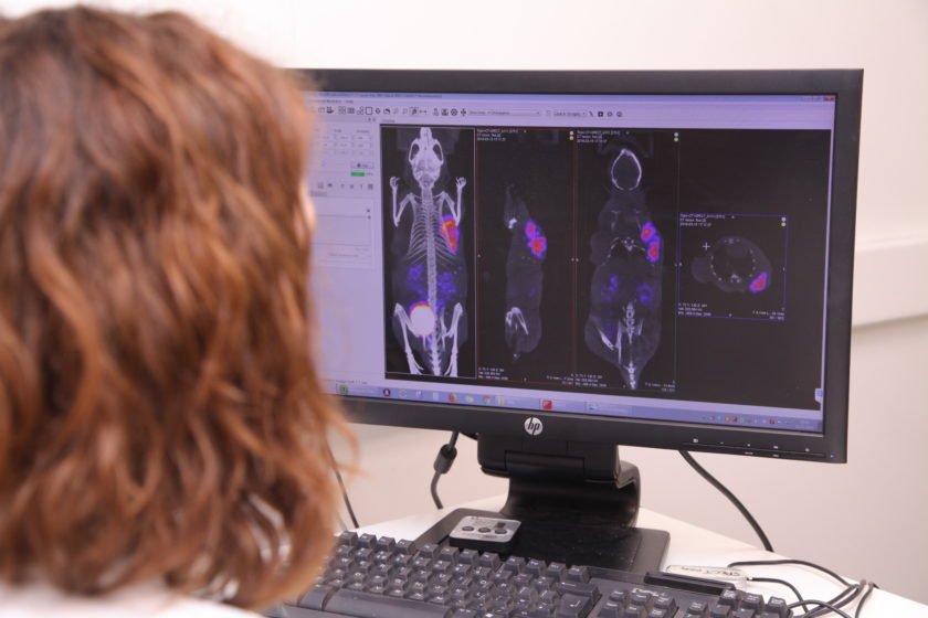
- This instrument allows single and multiple animal imaging (x4) for acquisition of 3D whole body datasets. We offer FDG and FLT PET imaging for detection of metabolic and proliferative changes of your model and have expert staff and a fully equipped radionuclide labelling laboratory to help you create targeted imaging agents for your research (both PET and SPECT agents).
- PET data is co-registered with the CT image to provide an attenuation and anatomical map for better quantitation and ease of analysis. CT may also be used alone to obtain high resolution images (to 10µm/pixel) for example to track bone and dentistry disease models or with a CT contrast agent to visualise vascular networks.
Bioscan NanoSPECT/CT (Single Photon Computed Tomography)
- Also offers single and multiple animal (x3) whole body 3D imaging, and is routinely used to image specific targets using radiolabelled ligands to sub-millimetre resolution. Anything from receptors upregulated in the area of interest or tumour, to the co-localisation of radiolabelled CAR-T cells or oncolytic viruses with tumours.
- An ECG gating system is available for cardiovascular applications and CT images may be acquired for co-registration with SPECT data.
We offer training to allow users to operate this instrument independently.
Bruker Icon 1T MRI (Magnetic Resonance Imaging)
- With excellent soft tissue contrast, our 1T MRI is an efficient screening tool for early detection and inclusion in study of some of the more difficult to detect orthotopic, spontaneous and metastatic cancer models. In full 3D acquisition mode it can be used to image details of interest for accurate volumetric measurements and has been used for anything from characterisation of tumour progression over time to detection and measurement of aneurysms to investigating the effects of brain trauma to cardiac imaging etc.
- With great ease of use, most users can be trained within a few hours to operate this instrument independently.
Perkin Elmer Lumina III Bioluminescence and Fluorescence imaging
- Our optical imaging system offers high throughput optical imaging (up to five mice imaged simultaneously in under 2 minutes). BLI may be used to track any cells bearing the reporter construct (luciferase) and high sensitivity means signal is detected even in the lowest quantities. With no “off target” signal, this modality is excellent for tracking changes in cell or disease burden.
- The FLI function offers a comprehensive range of excitation and emission filters so that variety of FLI probes may be used to target and image structures/processes of interest. The spectral un-mixing function minimises background signal and allows for multiple probes to be resolved when simultaneously imaged.
Again a very straight forward instrument to operate and training only takes a few hours.
Visual Sonics Vevo2100 Ultrasound
Our Vevo 2100 ultrasound combines good soft tissue resolution for anatomical details and measurements with the facility to investigate functional attributes such as blood flow.
- It may be used for internal tumour/structure detection and volumetric measurement, to calculate tumour vascularity, to investigate perfusion of vascular beds, to guide injections for accuracy etc. We are currently piloting an elastography study to investigate changes in tissue elasticity with disease.
Data Analysis
- Each instrument is equipped with its own easy-to-use acquisition software. The majority of analysis (both quantitative and image presentation) is done using VivoQuant software (Invicro) and training can be provided in its use. There is a bookable dedicated image analysis computer available in the imaging office so that analysis may be done outside of the Pre-clinical Imaging Facility itself.
- For optical imaging, Living Image (Perkin Elmer) software is used for analysis.
