BCI PhD student makes thesis competition national final

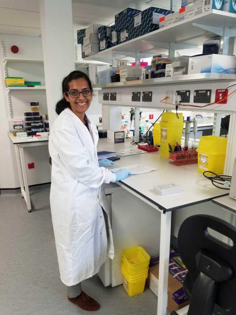
Congratulations to Vinaya Srirangam Nadhamuni, a Clinical Research Fellow at Barts Cancer Institute (BCI), Queen Mary University of London, who has progressed to the national final of the 2020 Vitae Three Minute Thesis® (3MT®) competition. As one of six finalists, Vinaya will be delivering a presentation about her research project live and online on Wednesday 16th September.
3MT® is an academic competition developed by the University of Queensland, Australia, that challenges PhD students to deliver a compelling spoken presentation on their research topic and its significance in just 3 minutes. Vinaya’s presentation is entitled ‘The war on cancer: understanding the immune system's strategies for fighting bowel cancer.’
As a histopathology trainee, Vinaya had completed 18 months of training in histopathology before pursuing a PhD as part of the Cancer Research UK City of London Centre Clinical Research Training Fellowship Programme. Vinaya is undertaking her research project in Professor Trevor Graham’s laboratory group in BCI’s Centre for Cancer Genomics and Computational Biology.
Ahead of the final, we spoke to Vinaya to find out more about her research and her experience in the competition.
What prompted you to enter the Vitae 3MT® competition? How have you found the experience so far?
I am often asked by my friends and family about the project I’m working on. My main aim in entering the competition was to come up with a short description that would explain this clearly (and maybe even wow them!). However, I have gained much more from this experience than I could have expected. Having to stop and think about the relevance of my work and how it fits into the bigger picture has been really useful, especially in terms of keeping me motivated to work even harder on my project.
Can you tell us a bit about your research project?
My project aims to understand how bowel cancer interacts with and escapes from the immune system. I am studying this by staining the different cell types in bowel cancer tissue with markers of different colours simultaneously and reviewing the stained tissue under the microscope. This will allow me to characterise the different immune cell types present- not only their quantity but also crucially their spatial interactions with each other and cancer cells.
This approach will help us identify mechanisms used by cancer cells to “escape” from the immune system. In turn, these insights will hopefully guide the development of treatments targeting these escape mechanisms. Eventually, this approach can be translated to a clinical setting such that we can determine the escape mechanisms present in a particular patient's cancer and provide targeted treatment, ultimately decreasing the significant mortality associated with bowel cancer.
From a histopathology point of view, a great deal of the clinical work in a pathology department involves looking at tissue under the microscope and using this information to diagnose cancer and identify risk factors which predict a worse prognosis. My project gives me the opportunity to take this process one step further by grouping the cells I see under the microscope into subtypes and understanding how they interact. I hope that this will deepen our understanding of the biological processes underlying the image that we see under the microscope.
How do you feel to have made it through to the 3MT® competition national final?
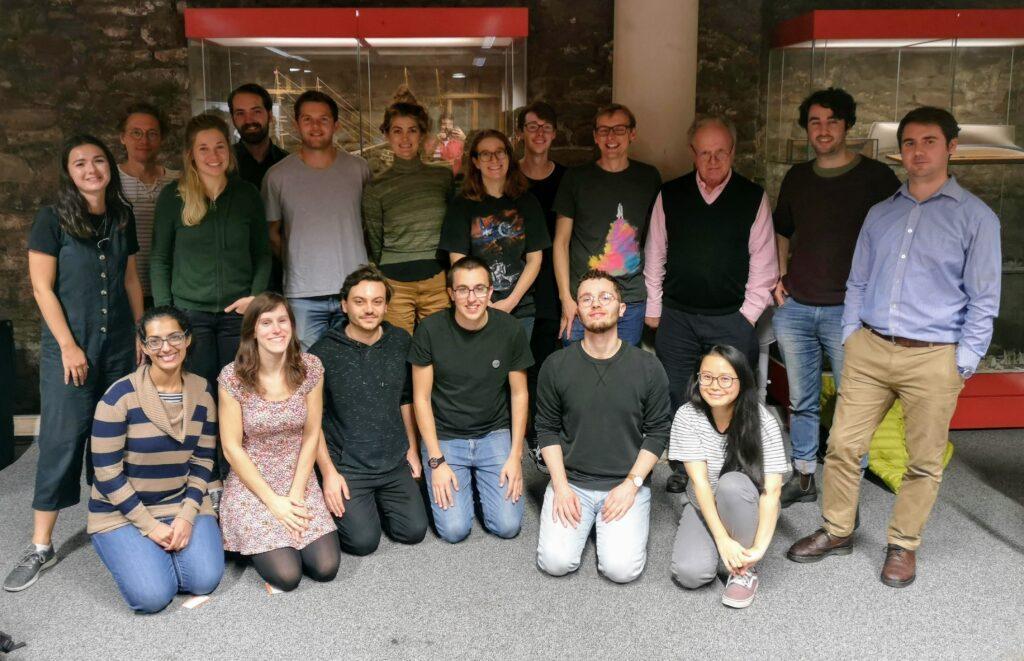
I am thrilled to have made it this far, especially considering 53 universities participated this year and only six finalists could be selected from the pool of candidates! I couldn’t have done it without the support of my fantastic lab. From helping me with my project at every turn to turning up to support me at the Queen Mary final, they have been absolutely amazing! I couldn’t ask for a more supportive team and am very grateful to have this opportunity to work with them during my PhD.

We wish Vinaya the best of luck in the final on 16th September. If you would like to watch the final and show your support, you can register online.
Category: Engagement, General News, Interviews

No comments yet