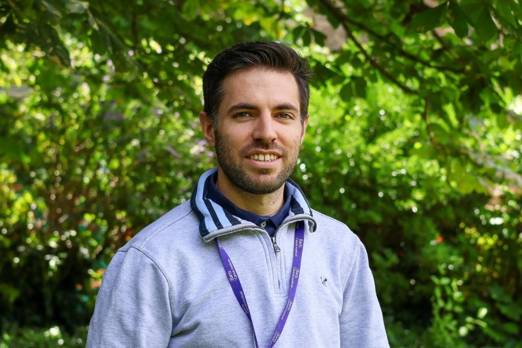
Introducing: Dr Oscar Maiques
We are pleased to welcome Dr Oscar Maiques to the Barts Cancer Institute (BCI) at Queen Mary University of London. Dr Maiques is establishing his first independent lab within our Centre for Cancer Biomarkers & Biotherapeutics. His research focuses on analysing medical images using advanced techniques to reveal how tissue structure changes in different tumours. Ultimately, these insights could help clinicians predict disease progression and tailor cancer treatments more effectively.
Dr Maiques currently has an exciting opportunity for a PhD student to join his lab. If you are interested in a career in cutting-edge cancer research, read more about the project and apply here.
Tell us about your journey into science.
I’ve loved science ever since high school. I was particularly fascinated by how biology works – everything from zoology to biochemistry. At that time, exciting advances were emerging in molecular biology, including new ways to manipulate DNA. I saw a huge potential for these new tools to help society, so I enrolled on a newly established Biotechnology degree at the University of Lleida in Catalonia, Spain, specialising in health science.
In my final year, I met Professor Xavier Matias-Guiu, a renowned cancer pathologist, who became my PhD supervisor. I shifted to a more clinical focus, studying how normal cells and tissue in a healthy person transform as cancer forms, and gaining experience in microscopy and pathology.

I learned a lot about the hallmarks of cancer and what was important for diagnosis. But I wanted to understand more about the mechanisms – why does cancer form and why do malignant cells leave their site and spread? During my PhD, I collaborated with Professor Victoria Sanz Moreno’s lab at King’s College London on a project where we provided tissue samples from human melanoma patients to her team for them to analyse biomarkers of metastasis. I felt that joining her lab as a postdoc would be the perfect next step, as it would enable me to use my understanding of clinical disease to ask crucial questions in their melanoma cell models.
When did you first come to the BCI?
Around this time, Vicky had begun to work more with human samples and adopt a more clinical focus. Moving to a dedicated translational cancer institute, like the BCI, where all of the tools and labs are focused on the same goal was therefore a natural transition. For me, it was perfect, and the microscope facility in particular was a game-changer. It gave me access to whole slide scanners, enabling us to develop a tool called multiplex immunohistochemistry that visualises tissue architecture across a whole slide using several different markers. The pathology facility was a fantastic help with developing and troubleshooting all the protocols.

What did your postdoctoral research at the BCI focus on?
My main area of expertise is the extracellular matrix (ECM) – the scaffolding network that maintains the 3D shape and structure of our tissues. I became interested in how the ECM is organised and how this organisation can allow cancer cells to migrate through it and move to other areas of the body (the process of metastasis). I have also been interested in how the type of ECM organisation can change the cytoskeleton of cells and make them more malignant and capable of spreading.
I believe that systematically quantifying these changes is important. Biology is complex, but clinicians need clear rules for stratifying and treating patients. I specialised in using image analysis to quantify how tissue architecture changes in normal and cancerous tissue, aiming to create tools to inform cancer diagnosis or predict recurrence.
In 2023, Vicky and her lab moved to the Institute of Cancer Research and I followed until I had the opportunity to return to the BCI as a group leader.

Why did you apply to be a group leader at the BCI?
Vicky has been very supportive of my goals to progress into a group leader position and when the position at the BCI came up she encouraged me to apply. These things happen once in life and it was a great career opportunity!
I think the BCI is a great place to build a stable career – the fact that so many people stay here for a long time speaks volumes. Another positive is that people already know me and what I’m capable of, and I have built several great collaborations here, such as with Kebs (Professor Kairbaan Hodivala-Dilke), Professor Tyson Sharp and Dr Oliver Pearce.
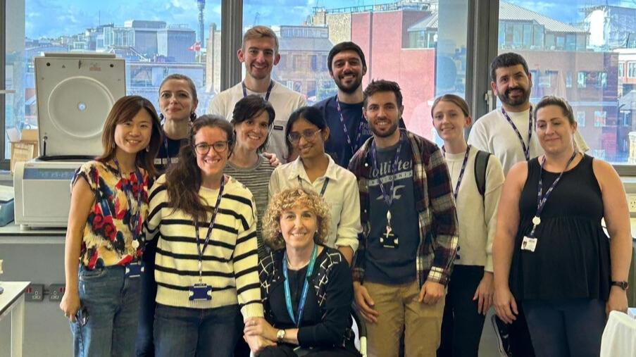
I’ve also become part of a great collaborative group with Professor Claude Chelala and Dr Vivek Singh within the Centre for Cancer Biomarkers and Biotherapeutics, who are also focused on using data-driven approaches to improve cancer prognosis and care. We have a multidisciplinary approach – we each have different areas of expertise and skill sets but can work together as a team with a common goal. This allows us to combine different perspectives and resources to tackle the same questions from different angles and more effectively help patients.
What will your lab focus on at the BCI?
I will build on what I was focusing on as a postdoc – exploring metastasis and how the tumour microenvironment and extracellular matrix impact it. I believe that the key to changing patients’ lives is understanding the earliest stages of malignancy and finding ways to diagnose and treat cancer early. I aim to find biomarkers that indicate whether a patient will develop cancer, whether it will metastasise, or whether the cells don’t pose a risk and we can spare the patient treatment.
We will use image analysis, tissue samples from patients, omics data and AI to generate the numbers clinicians need to make their treatment decisions. One unique aspect of our approach will be applying AI image analysis techniques to understand changes in the tumour microenvironment. Ultimately, we hope to reduce the need for invasive biopsies by using non-invasive imaging approaches to infer information about the molecular makeup of a tumour and inform treatment.
And finally: when you're not immersed in your research, how do you enjoy spending your time?
I love cycling and exploring new landscapes, particularly in Majorca. But most importantly, my wife and I have just had our first child, which is of course a big focus right now!
Category: General News, Interviews

No comments yet