Shining a Spotlight on AI Research at the Barts Cancer Institute
This year’s Nobel prizes in chemistry and physics recognised innovations in the field of artificial intelligence (AI). It’s an acknowledgement of the seemingly unstoppable rise in interest in this technology and its growing impact on society. Cancer research is no exception to this trend: AI is providing exciting new ways to understand, diagnose and predict the path of patients’ disease, offering opportunities to improve care and increase survival. At the Barts Cancer Institute (BCI), Queen Mary University of London, many of our researchers are using and improving these tools.
In this article, Bernhard Finke explores Nobel-Prize-winning research and the theory behind AI, and speaks with Dr Vivek Singh, Dr Oscar Maiques and Professor Claude Chelala, three researchers using cutting-edge AI in their work at the BCI. Bernhard is a PhD student in Dr Jun Wang’s lab, where he uses AI to build models that may one day help pathologists to more rapidly predict the course of disease in cancer patients.

The 2024 Nobel prizes in chemistry and physics celebrated very different advancements: one prize went to applied research published in the last decade, while the other went to foundational research carried out over 40 years ago. What they had in common was their celebration of the transformative power of AI.
Half of this year’s chemistry prize went to the creators of an AI-powered tool, which represents a breakthrough in the complicated problem of predicting the three-dimensional structure of proteins. Determining how a given protein will fold, which usually requires expensive experimental work, allows researchers to better understand their function. The tool, AlphaFold, is already massively helping to streamline the development of potential new cancer drugs.
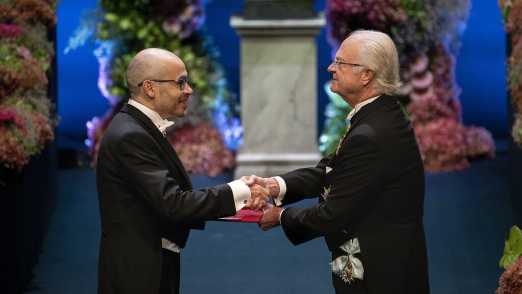
The physics prize was shared between Professor Geoffrey Hinton and Professor John Hopfield for their research into the progenitors of modern neural networks, a kind of AI algorithm that has formed the backbone of the current AI revolution. The networks developed by Hopfield and Hinton were loosely inspired by a simplified understanding of the similar networks of neurons found in human brains.
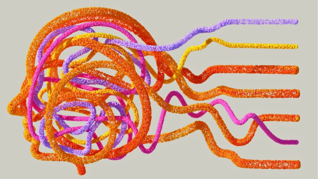
Like their biological counterpart, artificial neural networks process and transmit information from neuron to neuron. When fed with data, the networks automatically adjust the strengths of connections between different neurons: we call this process ‘learning’.
In my own research, for example, I feed a neural network with digital pathology data – microscopic images of cancer tissue, each labelled to indicate whether the tumour has spread, or ‘metastasised’. As the network sees more and more of these images, it learns and slowly becomes better at predicting whether a specific tumour is likely to metastasise.
Though there have been continual improvements in the algorithms used to help neural networks learn, a key factor that has precipitated the meteoric rise in AI research in recent years has been a combination of much greater data availability and more sophisticated hardware, which are both required to train ever larger neural networks. In medical fields like cancer research, obtaining large datasets, like those required to train an AI, can be a difficult task due to the rules around sharing sensitive patient data and the high cost of generating high-quality data. Researchers at the BCI are working to overcome these challenges and harness the power of AI to advance our understanding of oncology and improve outcomes for patients.
AI research at the BCI: Q&A with Dr Vivek Singh and Dr Oscar Maiques
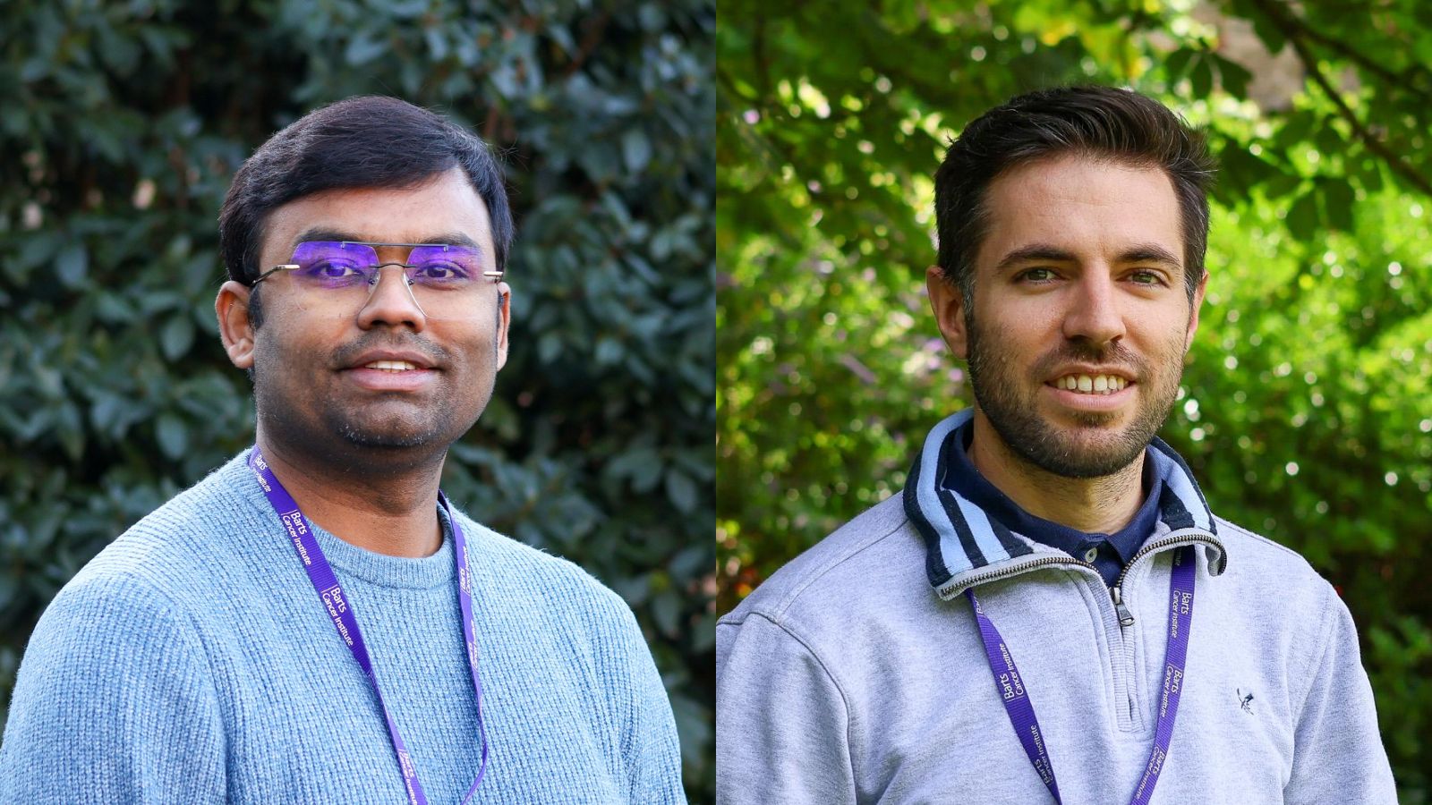
Dr Vivek Singh and Dr Oscar Maiques were both recruited to the BCI in the past year as part of the Barts Charity Precision Health Programme led by Professors Claude Chelala and Carol Dezateux, focusing on electronic health records, digital pathology, radiology, artificial intelligence and molecular datasets (‘omics’ data) to carry out research for patient and public benefit. Dr Singh and Dr Maiques will leverage their knowledge of AI technologies in the field of cancer research. I sat down with them both to hear more about their work and the opportunities and challenges that AI presents.
Tell me about the focus of your research at the BCI
Vivek: I specialise in applying AI and computer vision in cancer research, focusing on integrating radiology and digital pathology imaging with genomic data to improve early diagnosis of cancers such as breast, pancreas or colon. My work aims to enhance diagnostic accuracy, save time, and optimise resource utilisation in healthcare systems like the NHS, ultimately benefiting patient care.
Oscar: I use a variety of computational techniques, including AI, to analyze tumour tissue images, with the aim to predict metastasis and diagnosis in a variety of cancers. This will allow more precise clinical treatment and reduce the need for more invasive screening.
Could you describe an interesting AI research project you have undertaken recently?
Oscar: I’m working on an interesting collaboration with Professor John Marshall at the BCI to better understand the complicated mix of cells in pancreatic cancer. John is working with very complex data, which contain lots of different components in the tissue, including not only tumour cells but also healthy cells such as immune cells.
Using deep learning, we can determine the type of each cell, how it interacts with other neighbourhoods in the tissue, and how these factors relate to the patient’s prognosis. Ten years ago, doing this for a few patients would have taken months. But now we can analyse a cohort of 400 patients quickly and, theoretically, use this information to predict prognosis.
Vivek: My research focuses on using AI to speed up diagnosis in breast cancer and reduce turnaround time. I want to look at cases where a medical practitioner cannot easily determine whether a mass is benign from a scan and use AI to categorise it as benign or malignant to aid diagnosis. I am also looking at predicting molecular changes in a tumour using imaging, which will allow doctors to tailor care.
I am also working with radiologists at Barts Health NHS Trust, using AI large-language models (LLM) to generate automatic reports for imaging modalities. The LLM receives a medical image (like an X-ray or a CT scan) as input, analyses it, and directly generates a human-readable report summarising its findings. These reports are designed to assist doctors by reducing the time spent on routine documentation and allowing them to focus on complex cases and patient care.
What are the biggest challenges of applying AI to cancer research in your opinion?
Vivek: AI models need large amounts of data and we have to balance having large numbers of samples with building affordable technology that can actually be deployed in the NHS and have an impact. At Queen Mary University of London and the BCI, we have great computational resources, although it’s still expensive for us to house the data. Other institutions, like hospitals, don’t have the same computational infrastructure, which means that we need to adjust our models to work within these lower-resource environments. We’re currently in discussions to figure out the best methods, using a cloud-based infrastructure.
"Training with more diverse datasets allows
us to build models which are better suited
to groups that are often disadvantaged and underrepresented."
We’re also looking into further collaborations, so we have more data in diverse populations. Training with more diverse datasets allows us to build models which are better suited to groups that are often disadvantaged and underrepresented. Also, we need to be able to validate our models in other countries where they use different protocols and different machines. To be able to trust that our models really work, we need to show that they generalise to different data sources and types.
How do you see AI improving cancer treatment and diagnosis in the next few years?
Oscar: In my opinion, we won't have a single AI that can solve all of our problems in cancer research, as in other domains. Instead, there will be lots of different models trained to address individual problems. As each subtype of cancer is so distinct, we will also need different models for different types of cancers.
In the radiology field, we could see AI screening and early diagnosis tools used in clinical settings. In the future, we’ll be able to predict things like relapse, treatment response and molecular subtype and hopefully reduce the need for invasive measures (such as biopsies). These advances are more likely in cancers like breast, prostate and colorectal, but it’s probable that they will also be applied to many other tumours.
Vivek: I also want to add that AI has the potential to reduce the cost of regular testing by automating routine tasks and reducing the need for further testing. Globally, lots of countries don’t have enough experts and infrastructure, so we hope we can help to empower them using AI solutions. Our goal is to make cancer diagnosis faster, simpler, and more precise for everyone.
Looking to the future of AI in cancer research: Professor Claude Chelala
Professor Claude Chelala is a senior researcher at the BCI, specialising in computational genomics and computational data, whose recent work has branched out into AI. She specialises in breast and pancreatic cancer and oversaw the recruitment of Dr Vivek Singh and Dr Oscar Maiques.
Why it is such an important time to bring AI and cancer research together?
Claude: Cancer research advancements are producing an immense volume of multimodal data that must be meticulously analysed to inform diagnosis, prognosis and treatment. However, the field faces challenges in processing, analysing, storing, and reporting this data efficiently to fully unlock its potential. By addressing these needs, AI will accelerate novel cancer discoveries and its adoption within the NHS for enabling more precise diagnosis, personalised treatment plans, and improved patient outcomes.
The need and future potential for AI in cancer research are clear. Professor Dezateux and I successfully co-led the Precision Health research team based at Queen Mary University of London bringing AI and analytical skills to our University and strengthening our partnership with Barts Health NHS Trust. Since September 2022, this team has published more than 50 papers in peer-reviewed journals and contributed to leveraging over £16M of external research funding with more opportunities underway.
Category: General News, Interviews

No comments yet